From Images to Insights: Understanding Medical Image Processing
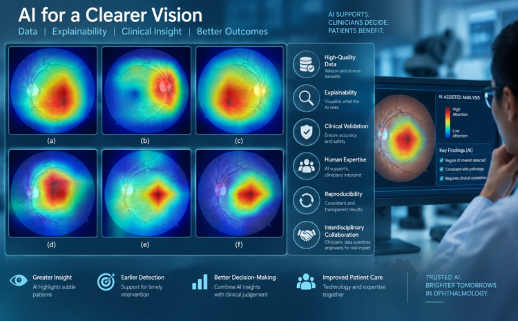
Learn how preprocessing, enhancement, segmentation, and analysis help transform raw ophthalmic images into useful information for AI and clinical research.
Medical images contain enormous amounts of information, but raw images are not always ready for direct analysis. Variations in image quality, illumination, noise, resolution, and acquisition techniques can affect how effectively an AI system interprets them.
Medical image processing provides a range of techniques for preparing and analysing these images. Preprocessing can improve image quality, while enhancement and segmentation can help isolate important anatomical structures or regions of interest.
For ophthalmic imaging, these processes can be particularly valuable when working with retinal photographs, OCT scans, and other forms of ocular imaging.
Understanding the journey from raw image → processed image → meaningful features → AI model is essential for anyone working at the intersection of ophthalmology and artificial intelligence.
METARES 3.0 – AVISHKAR provides participants with practical exposure to these concepts through hands-on learning and expert-led sessions.
